
")
MCS


The MCS is especially designed for:
- Large-scale, high-resolution, deep-tissue mapping
- Multispectral, high-speed, functional optical imaging
- Photostimulation while imaging through the same optical pathway (“photostimaging”)
- Two-photon microscopy and concurrent electrophysiology with computer-controlled placement of electrodes by Sutter micromanipulators
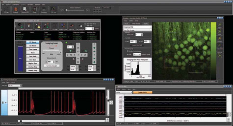
The MOM Computer System (MCS) is the software package MScan. This program has been designed to seamlessly control two-photon imaging, photostimulation and electrophysiology. While designed exclusively for use with the MOM, it is also compatible with other 2-photon platforms. MCS is designed to take on complex experiments in deep-tissue intravital imaging. Its intuitive user interface, is easy to use. The MCS and MOM together form a formidable tool to understand the most complex issues in neuroscience, immunology or oncology. Importantly, you will find in MCS the same standard of technical excellence that is the hallmark of all Sutter Instrument products.
The MScan software has been developed to simplify the many tasks inherent in a complicated imaging experiment. MScan is extensively multithreaded to take advantage of multicore processors. This ensures reliability and user interface responsiveness. Furthermore, MScan is multiuser based to facilitate sharing of a MOM microscope with MCS among experimenters. Experimenters can then send their data to other workstations for analysis. The MCS analysis program MView is available for free download on the Sutter Instrument website.
MCS includes a Windows workstation, National Instruments data acquisition boards, a Firewire CCD camera and a USB controlled MPC-200. The National Instruments boards included are a PCI-6110 board for imaging, a PCIe-6353 board for control of imaging and photostimulation laser power and a PCIe-6321 board for electrophysiology. The package is a turnkey system as all data acquisition boards and software come installed within the workstation.
An important feature provided in MScan is the ability to do bidirectional frame scanning with sub-pixel line offset adjustment. Conventional two-photon frame scanning has involved unidirectional scanning. In these scenarios, data is only recorded when sweeping in one direction across the sample. To increase the rate of data acquisition, it is then necessary to steer the laser beam back to the origin of the scan as quickly as possible to begin the subsequent line. As galvanometric scanners are most taxed and most likely to be damaged during these high-frequency movements, bidirectional scanning both increases the speed at which frames can be recorded and decreases the likelihood of damage to expensive galvanometers.
Imaging Features
4 imaging channels with independent gains and user-adjustable pixel duration
Bidirectional line scan with sub-pixel line offset adjustment
User or TTL triggered
Imaging modes
- XY movie
Real-time continuous zoom and rotation, 1° increment
XZ movie
Requires z-piezo nanopositioner - Timelapse
- Stack
Synchronized z focus and power modulation (linear or exponential)
On-line averaging - Fast stack
Fast XYZT time series when used with z- piezo nanopositioners
Synchronized z focus and power modulation (linear or exponential) - Line scan
User-drawn trajectory with arbitrary orientation and position - Region scan
User-designated collections of points, lines, rectangles, ellipses or polygons
Unlimited number of regions - Photostimulation scan
User-designated collections of points, lines, rectangles, ellipses or polygons
For each region, users can set: - Dwell time per pixel
- Laser intensity
- Duty cycle to output trains of light in the region
Control on imaging power
Dedicated fast analog output (submicrosecond response time) to control
laser intensity via Pockels cell allowing beam blanking on scan turn about.
Photostimulation
Dedicated fast analog output (submicrosecond response time) to control laser intensity via: - Pockels cell
Focus Control
Full XYZ control of objective placement with Sutter Instrument MPC-200
Photostimaging
Photostimaging is photostimulation during imaging. Photostimaging can be enabled during an XY movie, line scan or region scan.
Photostimaging requires a photostimulation laser (i.e., an ultrafast laser coupled to a Pockels cell or an analog-controlled bluegreen diode laser) sharing the imaging pathway with the imaging laser via a dichroic mirror.
Power measurement
interfaces with an optical laser power meter for accurate, real-time measurement of laser power to the preparation
3D map window
Stores multispectral frames or stacks in a 25 × 25 × 25 mm 3-D world in objective coordinates
Functional imaging
Real-time display of averaged intensities of regions of interest (ROIs) in scrolling plot
- ROIs can be rectangles, ellipses orpolygons
- Unlimited number of regions
- Automatic selection of the same region in other channels (useful for FRET)
Analog Inputs
8 analog channels, up to 250 kHz continuous acquisition rate
Software stimulator
Ideal to interface electrical or optogenetic stimulators
- 2 analog out channels
- 8 digital out lines
Targeted Patch-Clamping
Support for one Sutter Instrument micromanipulator
Control of two more micromanipulators with additional MPC-200 controller
Cellular Amplifier Control
Automatic real-time scaling of inputs via telegraph gain
Supports Axon Instruments AxoClamp 900A, Axopatch 200B, Multiclamp 700B, and, of course Sutter Instrument patch clamp amplifiers such as the dPatch, the IPA, and the dendrite.
Integrated development environment
Rich object model to control hardware, fully compatible with ActiveX Automation
MCS data files
Designed with GLP (Good Laboratory Practice) compliant header, visible from Windows shell
Contain imaging data, analog data and snapshots from CCD focusing camera
Standard tags and custom tags, queriable from other programs via ActiveX
Automation interface
Data file management
Semi-automatic backup of data files at the end of a session at up to two different locations (i.e., thumb drive, network disk…)
Bloggable lab notebook
Automatic log of events accessible via the built-in web browser.
Specifications
MCS
Dimensions
CPU 52 cm × 21 cm × 51 cm
Monitor 67 cm × 24 cm × 48 cm
Weight
CPU 14 kg
Monitor 7 kg
Electrical
115/230 Volts 50/60 Hz power line
Ordering information
MCS SYSTEM
System includes preinstalled MScan software, Windows workstation, data acquisition boards, Firewire CCD camera
The MCS system must be used with MOM systems equipped with MOM-DAQ, MPC-200 and ROE-200
MCS COMPONENTS
MOM-DAQ MOM data acquisition system (includes NI 6110E)
MPC-200-ROE Includes MPC-200 contoller and ROE-200
Note: The MOM-MCS communicates with the MPC-20o controller via the USB port for control of X, Y and Z movement. It is not compatible with the MP-285 controller.
Also See:
![]()
MOM
Product Flyer (PDF)
Related Products:
MOM
Two Photon
Movable Objective Microscope
3P-MOM
Threephoton Microscope

Beam Paths of the MOM
Video
(Youtube)